‘Listening to light’ to transform diagnosis of skin cancer

skinca

Scientists are developing a new photonics device that listens to light and could be capable of detecting skin cancer and other diseases more accurately than ever before, eliminating the need for unnecessary and invasive biopsies.

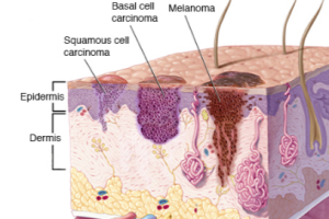
With around 232,000 people around the world, estimated to have been diagnosed with malignant melanoma in 2012, and with 55,500 deaths, early diagnosis of the disease could see hundreds of thousands of lives saved over the next ten years, improve quality of life and reduce healthcare costs.

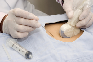
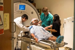
Traditionally, skin diseases are diagnosed visually by a physician using the naked eye or a magnifying glass and personal experience to make a decision. Invasive, uncomfortable, and potentially damaging procedures such as biopsies are often performed to confirm or exclude the presence of disease. This new breakthrough would give physicians an accurate and reliable way to objectively identify serious skin diseases for the first time.

“We are essentially listening to light, allowing us to see not just structures but molecules and biology on and under the skin, at depths and contrast never visualized before. It will enable physicians to make accurate and objective diagnosis of skin conditions for the first time.” said Professor Vasilis Ntziachristos, INNODERM Coordinator and Chair for Biological Imaging at the Technical University of Munich.

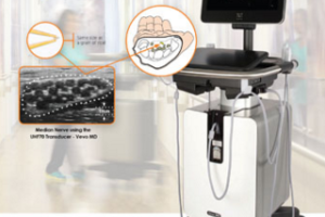
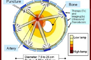
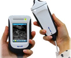
The method uses opto-acoustics, sending light waves of different wavelengths into the skin and detecting ultrasound waves generated within tissue in response to light absorption to build up an image of the skin tissue and specific molecules therein.

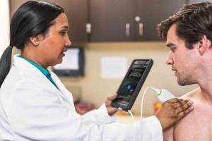
The prototype can visualize at depths up to 5mm under the skin, measures 4cm x 4cm x 7cm, no bigger than a small apple and can be placed on the skin to generate a high-resolution image in less than a minute. Being portable and of small form factor means that it could be used on expeditions or in remote areas of the world where a young doctor with little experience can make accurate, objective diagnoses.

“The device allows us to see blood vessels, skin oxygenation and potentially several novel pathophysiological features which are an integral area in the development of diseases. No one has ever been able to see like this before.” continued Professor Ntziachristos.

INNODERM, or Innovative Dermatology Healthcare based on Label-Free Spectral Optoacoustic Mesoscopy , combines the expertise of world-class engineers, scientists and clinicians in a consortium comprising 5 partners from 4 European countries.

The project has been awarded a grant of €3.8 million from Horizon 2020, the EU framework programme for research and innovation under the Photonics21 Public Private Partnership.

For more information: http://www.photonics21.org/index.php

Facebooktwittergoogle_plusredditlinkedinmail

New photonics technique to eliminate unnecessary thyroid cancer surgery

thyroid

A team of experts from around Europe has come together to develop a portable device with a hand-held probe that will dramatically reduce invasive diagnostic and therapeutic procedures in the treatment of thyroid nodules, saving approximately €450 million Euros every year.

The Photonics PPP and EU-funded Laser and Ultrasound Co-Analyzer for Thyroid Nodules, or LUCA, is a state-of-the-art device built to make thyroid nodule diagnosis more accurate and more objective.

With as much as 30 percent of adults in Europe, or 128.9 million people having to deal with a thyroid nodule at some point in their lives, accurate diagnosis has never been so important.

Each year in Europe alone, around 800,000 cases of detected thyroid nodules will be non-diagnostic, or indeterminate. Of all these cases, 150,000, or nearly 19%, will end up being benign and could have avoided surgery altogether.

At €3000 per operation, excluding additional medical costs, 150,000 unnecessary surgeries could mean saving over €450 million annually. “This money is wasted. We cannot, however, put a price on the wellbeing of a patient who does not have to undergo unnecessary surgery”, said ICREA Professor at ICFO- The Institute of Photonic Sciences, and the scientific coordinator of LUCA, Turgut Durduran.

According to Dr. Mireia Mora from the August Pi i Sunyer Biomedical Research Institute (IDIBAPS) in Barcelona,

“Current technology does not allow us to know whether a nodule is malignant or benign, before surgery takes place. We cannot take the risk of a misdiagnosis, so we operate.”

“LUCA will eliminate a lot of this guesswork. It will provide objective information so that we can see if a nodule is malignant or benign.”

Small in size, similar to a fizzy drink bottle, the LUCA probe is placed on the neck of the patient, sending light and ultrasound of different wavelengths and frequencies into the skin. “Ultrasound sees the structure and light sees the physiology, meaning we can see in much more detail than ever before”, Dr Mora said.

According to, Professor Durduran, “The LUCA platform combines ultrasound and near-infrared diffuse optical technologies in a single device and a probe. By combining information about tissue hemodynamics, chemical constitution as well as anatomy, the technique used by this device will overcome the shortcomings of present techniques while screening for malignant thyroid nodules.”

Women tend to be more affected by this condition. Out of 30% of Europeans who have thyroid nodules, women are three times more likely to develop nodules than men. However, of those diagnosed with a malignant nodule, the ratio then drops to 2:1, men to women, respectively, albeit the fact that thyroid cancer is still more prevalent in women than in men.

The implications of the LUCA device are extremely promising since it will not only signify a change in thyroid cancer screening techniques, but it may also have a potential use in the diagnosis of other cancers, such as the breast or any part of the body that is accessible.

Facebooktwittergoogle_plusredditlinkedinmail

New Certificate: Interventional Pain and MSK Ultrasound

interventional pain

ASRA will offer a new certificate in Interventional Pain and MSK Ultrasound to qualified physicians beginning in 2017.

The ASRA Board recently approved the development of the certificate, which will enable physicians to document skills and training in this highly specialized field of pain management. A committee will convene to develop the certificate program, which will require candidates to complete a written and practical exam as well as provide evidence of prior training and experience.


“The certificate helps ensure that a practitioner is properly trained and competent not only in the technical skills but the also in the judgment and communication skills that are essential to these procedures.” — Philip Peng, MBBS, FRCPC, Founder (Pain Medicine)


Ultrasound-guided pain procedures and musculoskeletal (MSK) injections require a high level of skill and experience in order to be safely administered. Sonography images are subject to individual interpretation depending on one’s experiences and training, and there is no formal training on Interventional Pain and MSK ultrasound during residency and fellowship. As a result, most physicians learn the techniques through hands-on workshops and online educational activities. There is increasing pressure from payers, employers, and the public to ensure physicians are competent and well trained.

“As a leader in providing high-quality, scientifically based training in these areas, it was natural for ASRA to move in this direction,” said Board member Samer Narouze, MD, PhD, who has championed the program’s development.

Philip Peng, MBBS, FRCPC, Founder (Pain Medicine), co-developer and ASRA faculty member, said the certificate reflects more than just technical proficiency.

“ASRA educational programs are well-respected, and the quality is well known in the field,” Peng said. “The certificate helps ensure that a practitioner is properly trained and competent not only in the technical skills but the also in the judgment and communication skills that are essential to these procedures.”

Continue reading here

Facebooktwittergoogle_plusredditlinkedinmail

FUJIFILM VisualSonics launches first Ultra High Frequency clinical ultrasound imaging system

Vevo

FUJIFILM VisualSonics Inc., a world leader in ultra high frequency ultrasound imaging systems and subsidiary of FUJIFILM SonoSite, Inc., announces CE mark for the Vevo® MD, the world’s first Ultra High Frequency (UHF) clinical ultrasound system. With multiple successes in preclinical research over the last decade, today marks a major milestone for FUJIFILM VisualSonics as it expands into the clinical market.

“As the undisputed leader in ultra high frequency imaging systems, FUJIFILM VisualSonics once again advances the field of ultrasound with the launch of the Vevo MD,” said Masayuki Higuchi, president & CEO, FUJIFILM SonoSite, Inc. “We are proud to be the first to bring to market this exciting innovation that is sure to have high impact on the medical imaging community while expanding the product portfolio of FUJIFILM SonoSite.”

The Vevo MD is truly a unique ultrasound system, as it operates at much higher frequencies than any conventional ultrasound system currently available. Along with the Vevo MD system, FUJIFILM VisualSonics also introduces the UHF Series of transducers. This patented transducer technology is capable of operating in a range of frequencies up to 70 MHz, a tremendous increase in resolution compared to conventional ultrasound systems.

“The Vevo MD allows medical professionals to see what they have never seen before—unparalleled image resolution down to 30 micrometers. This is less than half the size of a grain of sand,” said Renaud Maloberti, vice president & general manager of FUJIFILM VisualSonics. “Imagine the great potential this technology has across unexplored applications to visualize the smallest, highly detailed anatomy in a way that has never been done before.”

“We believe this technology has a role to play in a range of clinical application areas from neonatology, vascular, musculoskeletal, dermatology, or other small parts that are within the first 3 cm of the body,” said Andrew Needles, director of marketing at FUJIFILM VisualSonics.“We also know that there are new and interesting areas to be discovered. The Vevo MD gives us and our users the opportunity to make those discoveries together.”

The Vevo MD is now commercially available with CE mark in the majority of European Union countries. For more information and contact details please visit www.vevomd.com or contact FUJIFILM VisualSonics at www.visualsonics.com.


Facebooktwittergoogle_plusredditlinkedinmail

Philips starts shipping Lumify in the U.S., an app-based ultrasound

Lumify


Amsterdam, the Netherlands – Royal Philips (NYSE: PHG, AEX: PHIA) announced that Lumify, a smart device ultrasound solution, is available for purchase by licensed healthcare providers or organizations in the U.S. Lumify is offered as part of a novel subscription model unique to the industry. From Philips’ online portal, users will be able to order transducers, manage flexible subscriptions and access Philips’ support, training and IT services through the new offering. Philips’ new ultrasound digital health approach connects off-the-shelf compatible smart devices, a mobile application, advanced ultrasound transducer technology, integrated IT and support services to help healthcare providers improve patient care and reduce costs.

Since debuting the smart device ultrasound concept in 2014, Philips has enhanced the solution for more clinical uses including acute care and office practice. As a comprehensive solution, Lumify extends the benefits of ultrasound to more places by leveraging cloud-enabled and tablet technology, and bringing connectivity, flexibility and mobility into the hands of more healthcare providers.

Continue reading here

Facebooktwittergoogle_plusredditlinkedinmail

DBAC Ultrasound Cuff to Stop Bleeding

DBAC ultrasound

Since 2008, Siemens and University of Washington have been working on a very ambitious project. DBAC (Deep Bleeder Acoustic Coagulation). Funded by DARPA (Defense Advanced Research Project Agency), the goal is to use HIFU (High Intensity Focused Ultrasound) to stop bleeding in deep vessels.  Here they report the latest findings in this two part paper.

Part I: development and in vitro testing of a research prototype cuff system

Part II: in vivo testing of a research prototype system


Facebooktwittergoogle_plusredditlinkedinmail

Handheld ultrasound Signostics receives $35 million from private investor KKR

handheld ultrasound


CLOVELLY PARK, SOUTH AUSTRALIA (November 16, 2015) – Signostics, a start-up innovator in the field of highly miniaturized ultrasound technology, today announced a private investment of $35 million dollars from KKR, a leading global investment firm.

Since launching its first product in 2009, Signostics has remained focused on the development of handheld ultrasound technology. The new funding by KKR will allow Signostics to pursue cutting-edge medical innovations and to expand and accelerate global marketing and distribution for new products. Prior to this transaction, Signostics re-domiciled from Australia to become a Delaware-based corporation with newly-established headquarters in Kirkland, Washington.

“KKR offers us the privilege of working with a highly regarded long-term capital partner which will enable us to create next-level innovations and beyond, aimed at greater disruption and democratization of the global ultrasound market,” said Kevin Goodwin, Chief Executive Officer of Signostics who joined the company earlier this year. “We feel very fortunate to have gained the support of KKR as we set out to build on our work in this field over the last decade.”

Continue reading here

Facebooktwittergoogle_plusredditlinkedinmail

World first: blood-brain barrier opened non-invasively to deliver chemotherapy

Blood-Brain Barrier

Toronto, ON (November 8, 2015) – Sunnybrook scientists made history this week as they used focused ultrasound to non-invasively breach the blood-brain barrier and more effectively deliver chemotherapy into the brain tumour of a patient.

Each person has a protective barrier that normally restricts the passage of substances from the bloodstream into the brain protecting it from toxic chemicals.

“The blood-brain barrier (BBB) has been a persistent obstacle to delivering valuable therapies to treat disease such as tumours,” says Dr. Todd Mainprize, principal investigator of the study and Neurosurgeon in the Hurvitz Brain Sciences Program at Sunnybrook Health Sciences Centre.

“We are encouraged that we were able to temporarily open this barrier in a patient to deliver chemotherapy directly to the brain tumour.”

The research team infused a chemotherapy drug, then tiny, microscopic bubbles, into the bloodstream of a patient with a malignant brain tumour. The microbubbles are smaller than red blood cells and pass harmlessly through the circulation. The researchers then used state-of-the-art MRI-guided focused low-intensity ultrasound (sound waves) to target blood vessels in the BBB area near the tumour. The waves repeatedly compress and expand the microbubbles, causing them to vibrate and loosen tight junctions of the cells comprising the BBB. Once the barrier was opened, the chemotherapy flowed through and deposited into the targeted regions.

“Some of the most exciting and novel therapeutics for the treatment of malignant brain tumours are not able to reach the tumour cells because of the blood brain barrier. This technique will open up new opportunities to deliver potentially much more effective treatments to the targeted areas,” says Dr. Todd Mainprize, also an Assistant Professor in the Division of Neurosurgery at University of Toronto.

Dr. Kullervo Hynynen, Director of Physical Sciences at Sunnybrook Research Institute, worked with industry partner Insightec for almost two decades to develop the technology and bring it to a clinic-ready state. “The success of this case is gratifying,” he says. “My hope now is that many patients will eventually benefit from it.”

Less than 24 hours after breaching the blood brain barrier, the tumour and some surrounding tissues were surgically removed and sent to pathology to measure differences in the concentration of chemotherapy that deposited in the area treated by the focused ultrasound and the area not treated.

“Breaching this barrier opens up a new frontier in treating brain disorders,” says Dr. Neal Kassell, chairman of the Focused Ultrasound Foundation. “We are encouraged by the momentum building for the use of focused ultrasound to non-invasively deliver therapies for a number of brain disorders.”

The success of this research opens up the potential for delivering drug therapies to parts of the brain protected by the blood brain barrier, including researching treatments for patients with various kinds of brain tumours, Alzheimer’s disease, and some psychiatric conditions.

“This is a very important step in the development of MR-guided focused ultrasound technology,” says Eyal Zadicario, VP R&D and Director of Neuro Programs at Insightec. “We continue to push the technology into new clinical applications that can have significant impact where it matters most – to patients.”

This case was the first of up to 10 participants in this study, which is testing for feasibility, safety and preliminary efficacy. The participants are those already scheduled for traditional neurosurgery to remove parts of their brain tumour.

This study is funded by the Focused Ultrasound Foundation. The research equipment is funded by the Canada Foundation for Innovation and Ontario Ministry of Research and Innovation, with support from the Bombardier Foundation. The research has also received significant support from the Canadian Institutes of Health Research and National Institutes of Health. Dr. Hynynen holds the Canada Research Chair in Imaging Systems and Image-Guided Therapeutics. The focused ultrasound apparatus and technology are Insightec’s Exablate Neuro System.

Media contact:

Nadia Norcia Radovini, nadia.radovini@sunnybrook.ca, (416) 480-4040

See original post and video here


Facebooktwittergoogle_plusredditlinkedinmail

Heat-activated ‘grenade’ to target cancer

Researchers have developed cancer drug-packed ‘grenades’ armed with heat sensitive triggers, allowing for treatment to be targeted directly at tumours, according to two studies due to be presented at the National Cancer Research Institute (NCRI) Cancer Conference in Liverpool.

The team based at The University of Manchester has been developing liposomes – small, bubble-like structures built out of cell membrane that are used as packages to deliver molecules into cells – to carry drugs into cancer cells. The challenge, as with any treatment, is to direct the liposomes and their payload directly to tumours while sparing healthy tissue.

… Read more here

Facebooktwittergoogle_plusredditlinkedinmail
1 2 3 4