Low Intensity Pulsed Ultrasound doesn’t improve bone fracture healing

broken legLow intensity ultrasound after surgical repair of a bone fracture is a popular treatment to improve recovery, but it doesn’t work, says a large international study led by researchers at McMaster University.

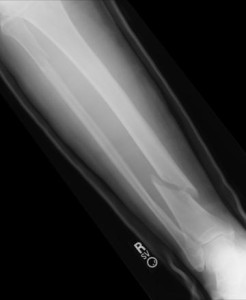
In a clinical trial published by The BMJ today, the researchers show no difference in the recovery time between using low intensity pulsed ultrasound (LIPUS) or a placebo device for patients with a fractured tibia (shinbone).

An accompanying editorial, also published today, suggests “it is time to abandon this ineffective treatment” now that there is strong evidence it doesn’t work.

“LIPUS is commonly used in North America to accelerate fracture healing — generating about $250 million in sales a year – but there has been no clear evidence of benefit to support its use,” said principal investigator Jason Busse.

Busse is an associate professor of anesthesia and a researcher with the Michael G. DeGroote Institute for Pain Research and Care at the Michael G. DeGroote School of Medicine at McMaster.

LIPUS was approved for fracture healing by the U.S. Food and Drug Administration (FDA) in 1994. Interventions to aid recovery have been popular for tibia fractures since this common fracture heals slowly and often needs further surgery to heal completely.

The international research team conducted a randomized controlled trial of 501 patients at 43 academic trauma centres in North America who underwent surgical repair for a tibia (lower leg) fracture between 2008 and 2012. The patients were assigned 20-minute daily treatment with either a LIPUS or a placebo device which looked the same. Everyone involved, including the physicians, data collectors, data analysts and the patients were blind to which treatment was used. Patients were followed until x-rays showed their fracture was healed, or for 12 months.

There was no difference in time to functional recovery whether patients were treated with the active or placebo device. There was also no difference in quality of life, return to work, leisure activities, or time to full weight-bearing.

Co-principal investigator Mohit Bhandari, who is a professor of surgery at McMaster, pointed out the importance of ensuring medical devices are supported by evidence.

“LIPUS was approved for fracture healing on the basis of small trials that had important limitations, and they focussed on radiographic healing instead of patient important outcomes,” said Bhandari, who also holds the Canada Research Chair in Evidence-Based Orthopaedic Surgery.

“The new trial results establish that LIPUS has no role in managing patients with surgically repaired fractures.”

In the accompanying editorial in The BMJ, Xavier Griffin, an associate professor of trauma surgery at the University of Oxford, said the results were clear.

“These authors report important patient-centred outcomes…showing that low intensity pulsed ultrasound is of no benefit to adults with tibia fractures,” he said. “It is time for us to make good use of their determination and abandon this ineffective treatment.”

This study was funded by the Canadian Institutes of Health Research and Smith & Nephew, specialists in surgical devices and wound care.

Original article can be found here.

[yop_poll id=”1″]

Facebooktwittergoogle_plusredditlinkedinmail

John Grisham’s book “The Tumor” skillfully mix fiction with science

john grishamThis book is free, with only 46 pages. An easy read, but with a powerful story and message. John Grisham writes on behalf of the whole Focused Ultrasound Foundation. For most of us working with ultrasound, it should spark that sense of urgency to get to the finish line and see your product helping those suffering from this terrible disease.

You can download John Grisham’s free book “The Tumor” here.


And in this TEDx talk, together with Dr. Neal Kassell,  John Grisham explains with good sense of humor how and why he got involved with the Focused Ultrasound Foundation:


Facebooktwittergoogle_plusredditlinkedinmail

‘Listening to light’ to transform diagnosis of skin cancer

skinca

Scientists are developing a new photonics device that listens to light and could be capable of detecting skin cancer and other diseases more accurately than ever before, eliminating the need for unnecessary and invasive biopsies.

With around 232,000 people around the world, estimated to have been diagnosed with malignant melanoma in 2012, and with 55,500 deaths, early diagnosis of the disease could see hundreds of thousands of lives saved over the next ten years, improve quality of life and reduce healthcare costs.

Traditionally, skin diseases are diagnosed visually by a physician using the naked eye or a magnifying glass and personal experience to make a decision. Invasive, uncomfortable, and potentially damaging procedures such as biopsies are often performed to confirm or exclude the presence of disease. This new breakthrough would give physicians an accurate and reliable way to objectively identify serious skin diseases for the first time.

“We are essentially listening to light, allowing us to see not just structures but molecules and biology on and under the skin, at depths and contrast never visualized before. It will enable physicians to make accurate and objective diagnosis of skin conditions for the first time.” said Professor Vasilis Ntziachristos, INNODERM Coordinator and Chair for Biological Imaging at the Technical University of Munich.

The method uses opto-acoustics, sending light waves of different wavelengths into the skin and detecting ultrasound waves generated within tissue in response to light absorption to build up an image of the skin tissue and specific molecules therein.

The prototype can visualize at depths up to 5mm under the skin, measures 4cm x 4cm x 7cm, no bigger than a small apple and can be placed on the skin to generate a high-resolution image in less than a minute. Being portable and of small form factor means that it could be used on expeditions or in remote areas of the world where a young doctor with little experience can make accurate, objective diagnoses.

“The device allows us to see blood vessels, skin oxygenation and potentially several novel pathophysiological features which are an integral area in the development of diseases. No one has ever been able to see like this before.” continued Professor Ntziachristos.

INNODERM, or Innovative Dermatology Healthcare based on Label-Free Spectral Optoacoustic Mesoscopy , combines the expertise of world-class engineers, scientists and clinicians in a consortium comprising 5 partners from 4 European countries.

The project has been awarded a grant of €3.8 million from Horizon 2020, the EU framework programme for research and innovation under the Photonics21 Public Private Partnership.

For more information: http://www.photonics21.org/index.php

Facebooktwittergoogle_plusredditlinkedinmail

New photonics technique to eliminate unnecessary thyroid cancer surgery

thyroid

A team of experts from around Europe has come together to develop a portable device with a hand-held probe that will dramatically reduce invasive diagnostic and therapeutic procedures in the treatment of thyroid nodules, saving approximately €450 million Euros every year.

The Photonics PPP and EU-funded Laser and Ultrasound Co-Analyzer for Thyroid Nodules, or LUCA, is a state-of-the-art device built to make thyroid nodule diagnosis more accurate and more objective.

With as much as 30 percent of adults in Europe, or 128.9 million people having to deal with a thyroid nodule at some point in their lives, accurate diagnosis has never been so important.

Each year in Europe alone, around 800,000 cases of detected thyroid nodules will be non-diagnostic, or indeterminate. Of all these cases, 150,000, or nearly 19%, will end up being benign and could have avoided surgery altogether.

At €3000 per operation, excluding additional medical costs, 150,000 unnecessary surgeries could mean saving over €450 million annually. “This money is wasted. We cannot, however, put a price on the wellbeing of a patient who does not have to undergo unnecessary surgery”, said ICREA Professor at ICFO- The Institute of Photonic Sciences, and the scientific coordinator of LUCA, Turgut Durduran.

According to Dr. Mireia Mora from the August Pi i Sunyer Biomedical Research Institute (IDIBAPS) in Barcelona,

“Current technology does not allow us to know whether a nodule is malignant or benign, before surgery takes place. We cannot take the risk of a misdiagnosis, so we operate.”

“LUCA will eliminate a lot of this guesswork. It will provide objective information so that we can see if a nodule is malignant or benign.”

Small in size, similar to a fizzy drink bottle, the LUCA probe is placed on the neck of the patient, sending light and ultrasound of different wavelengths and frequencies into the skin. “Ultrasound sees the structure and light sees the physiology, meaning we can see in much more detail than ever before”, Dr Mora said.

According to, Professor Durduran, “The LUCA platform combines ultrasound and near-infrared diffuse optical technologies in a single device and a probe. By combining information about tissue hemodynamics, chemical constitution as well as anatomy, the technique used by this device will overcome the shortcomings of present techniques while screening for malignant thyroid nodules.”

Women tend to be more affected by this condition. Out of 30% of Europeans who have thyroid nodules, women are three times more likely to develop nodules than men. However, of those diagnosed with a malignant nodule, the ratio then drops to 2:1, men to women, respectively, albeit the fact that thyroid cancer is still more prevalent in women than in men.

The implications of the LUCA device are extremely promising since it will not only signify a change in thyroid cancer screening techniques, but it may also have a potential use in the diagnosis of other cancers, such as the breast or any part of the body that is accessible.

Facebooktwittergoogle_plusredditlinkedinmail

New Certificate: Interventional Pain and MSK Ultrasound

interventional pain

ASRA will offer a new certificate in Interventional Pain and MSK Ultrasound to qualified physicians beginning in 2017.

The ASRA Board recently approved the development of the certificate, which will enable physicians to document skills and training in this highly specialized field of pain management. A committee will convene to develop the certificate program, which will require candidates to complete a written and practical exam as well as provide evidence of prior training and experience.


“The certificate helps ensure that a practitioner is properly trained and competent not only in the technical skills but the also in the judgment and communication skills that are essential to these procedures.” — Philip Peng, MBBS, FRCPC, Founder (Pain Medicine)


Ultrasound-guided pain procedures and musculoskeletal (MSK) injections require a high level of skill and experience in order to be safely administered. Sonography images are subject to individual interpretation depending on one’s experiences and training, and there is no formal training on Interventional Pain and MSK ultrasound during residency and fellowship. As a result, most physicians learn the techniques through hands-on workshops and online educational activities. There is increasing pressure from payers, employers, and the public to ensure physicians are competent and well trained.

“As a leader in providing high-quality, scientifically based training in these areas, it was natural for ASRA to move in this direction,” said Board member Samer Narouze, MD, PhD, who has championed the program’s development.

Philip Peng, MBBS, FRCPC, Founder (Pain Medicine), co-developer and ASRA faculty member, said the certificate reflects more than just technical proficiency.

“ASRA educational programs are well-respected, and the quality is well known in the field,” Peng said. “The certificate helps ensure that a practitioner is properly trained and competent not only in the technical skills but the also in the judgment and communication skills that are essential to these procedures.”

Continue reading here

Facebooktwittergoogle_plusredditlinkedinmail